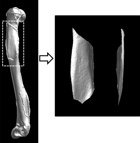
Fig. 6.

Using the three-dimensional reconstructed data, the cortical bone surface of the region of interest (the muscle/tendon attachment site) was extracted along the gel marker exposed on X-ray. The image on the right shows the deltoid attachment site on the humerus after trimming.
