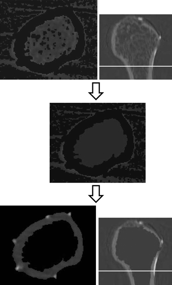
Fig. 7.

The top images show that the trabecular bones and bone marrow in the medullary cavity were not removed in the cross-section. The middle image indicates that the trabecular bones and bone marrow in the medullary cavity were assigned the value of air, only cortical bone remaining. The cross-section at the threshold separating the cortical bone and air is shown in the lowest image and indicates that trabecular bone or bone marrow was absent inside the bone. The right side images indicate the sagittal plane of the humerus and the white lines show the positions of the images to the left.
