
Figure 6.
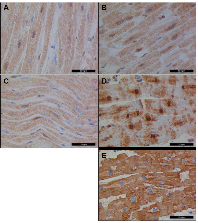
Immunohistochemistry was performed with a monoclonal antibody against GATAD1 on left ventricular tissue from four individuals. Normal tissue from a male (A) and a female (B) display nuclear staining of GATAD1 with a homogeneous striated pattern in the extranuclear space, similar to what was noted in tissue from an individual with DCM due to a mutation in TPM1 (C). In contrast, GATAD1 in biopsy tissue from the proband shows an abnormal staining pattern (D). While there is still nuclear localization, extranuclear distribution of GATAD1 is perturbed whereas actin staining of an adjacent section (E) was indistinguishable from normal controls (data not shown). In addition, the globular morphology of nuclei is unique from the spindle shape observed in control tissue.