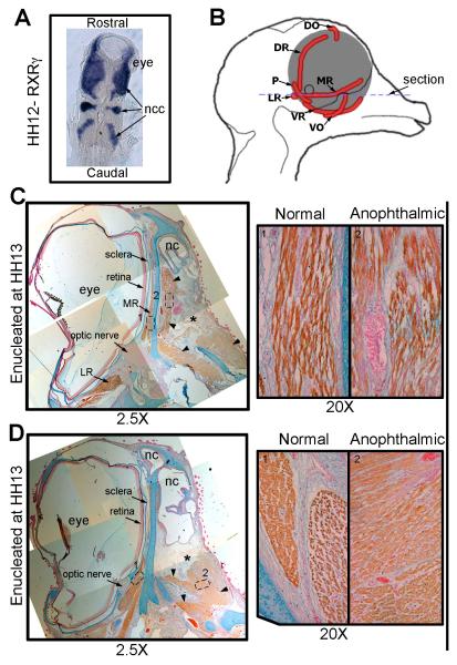
Figure 10. Surgical removal of the developing eye in the chick disrupts EOMs.
A) Dorsal view in situ hybridization for RXRγ on a HH stage 12 (~E2) chick embryo, showing the close contact between the developing eye and the neural crest cells (blue). B) Schematic representing the chick head of an E14 embryo seen from the midline showing the positioning of the EOMs (red) of the left eye. The blue dotted line suggests the position of the sections in C and D. (C and D) HH stage 40 (E14) chick embryo that was enucleated at HH stage 13 labeled for Myosin (brown), Alcian blue (cartilage- blue) and nuclear fast red (nuclei-red). The position where the eye would have developed is denoted by the *. No retina, optic nerve or sclera has developed in this region. However, extraocular muscles are seen (arrow heads). C) The medial rectus muscle on the anophthalmic side of the embryo (right) has become fully differentiated muscle but is misshapen and has a looser morphology compared to those seen on the control (left) side. D) The other EOMs show reduced organization (right) compared to the control side (left)and individual muscles are no longer discrete and identifiable. Ncc, neural crest cell; MR, medial rectus; VR, ventral rectus; LR, lateral rectus; VO, ventral oblique; P, pyramidalis; DR, dorsal rectus; DO, dorsal oblique; nc, nasal cavity.

