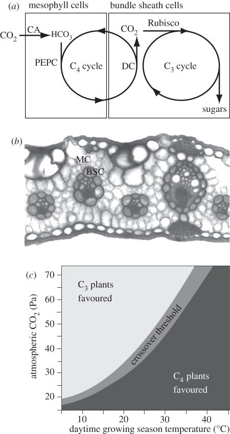
Figure 1.
Mechanism of C4 photosynthesis. (a) Simplified schematic of the C4 syndrome, showing how the C3 pathway is isolated from the atmosphere in most C4 species within a specialized tissue composed of bundle sheath cells (BSC). The C4 pathway captures CO2 within mesophyll cells (MC) using the enzymes carbonic anyhdrase (CA) and phosphoenolpyruvate carboxylase (PEPC). It then transports fixed carbon to the BSC where decarboxylase enzymes (DC) liberate CO2, which accumulates to high concentrations around Rubisco. (b) Transverse section of a C4 leaf, showing the arrangement of MC and BSC. In this picture, the BSC are thicker walled, are stained darker and form rings surrounding the veins (adapted from Watson & Dallwitz [9]). Enlarged BSC relative to MC and close vein spacing are typical of C4 leaves, and this pattern is referred to as ‘Kranz anatomy’. (c) Explanation of how variation in CO2 and temperature favours C3 or C4 species [10,11]. The schematic shows the range of CO2 partial pressures and temperatures predicted to favour the growth of either C3 or C4 species, based on the maximum quantum yield of photosynthesis. Quantum yield is a measure of photosynthetic efficiency, which declines in C3 plants as photorespiration increases at low CO2 and high temperature, (reproduced with permission from Edwards et al. [3], which was adapted from Ehleringer et al. [11]).

