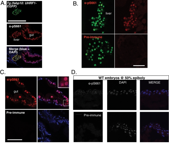
FIGURE 7:
Phosphorylated endogenous Uhrf1 is cytoplasmic. (A) α-pS661 detects both overexpressed and endogenous phosphorylated UHRF1 in zebrafish. Top, GFP signal in hepatocytes of Tg(fabp10:UHRF1-GFP); middle, immunofluorescence with α-pS661; bottom, merge with DAPI. The gut and liver are labeled. Magnification: 20×; scale bar: 250 μm. (B) Sections of 5-dpf zebrafish expressing GFP-tagged UHRF1 under the control of the hepatocyte specific promoter (Tg(fabp10:UHRF1-EGFP)). Left, GFP expression in hepatocytes; right, immunofluorescence with α-pS661 (top) or preimmune (bottom). Confocal images taken with identical parameters. Scale bar: 50 μm. (C) Left, immunofluorescence of enterocytes with α-pS661 or preimmune. Right, merge with DAPI. Inset, 63× magnification of endogenous phosphorylated UHRF1 in the zebrafish gut with predominant nuclear localization. Confocal images taken with identical parameters. Scale bar: 50 μm. (D) Endogenous UHRF1 is phosphorylated in early development as seen with immunofluorescence on cryosections at 50% epiboly. Bottom, left, immunofluorescence on cryosections of embryos at 50% epiboly with preimmune sera; middle, DAPI alone; right, merge. Top, left, immunofluorescence with α-pS661; middle, DAPI alone; right, merge. Magnification: 63×; scale bar: 25 μm.

