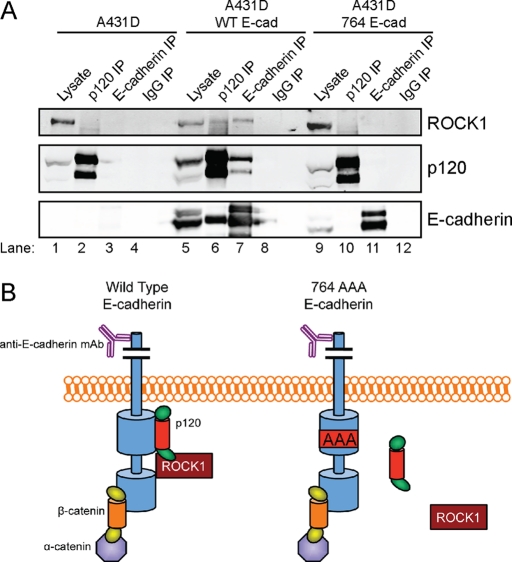
FIGURE 4:
ROCK1 physically associates with the cadherin complex in a p120-dependent manner. (A) Coimmunoprecipitation of ROCK1 with wild-type E-cadherin in DSP–cross-linked A431D cells. Western blot analysis of ROCK1, p120, and E-cadherin in whole-cell lysates containing 10% of input (lanes 1, 5, and 9) or immunoprecipitations generated using p120 (lanes 2, 6, and 10), E-cadherin (lanes 3, 7, and 11), or control IgG (lanes 4, 8, and 12) mAbs. (B) A model depicting p120-dependent association of ROCK1 with the cadherin complex, as detected by immunoprecipitation in (A). The extracellular domain of E-cadherin has been truncated in this schematic.

