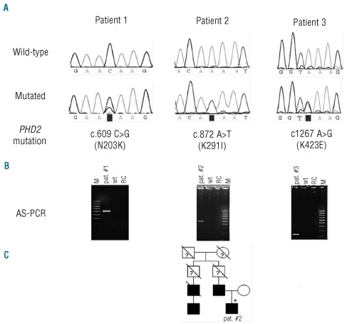
Figure 1.
Mutational analysis results and localization of the PHD2 mutations. (A) Sequencing results of wild-type and mutated alleles in 3 patients. Nucleotide positions (GenBank accession, NM 022051), nucleotide changes and corresponding amino acid changes are indicated below. (B) Confirmation of the new mutations by allele-specific PCR (AS-PCR) and agarose gel electrophoresis. AS-PCR fragment lengths agree with the respective primer positions (Online Supplementary Table S1). Lane M: molecular weight marker, lane pat #: patient’s DNA amplified with mutation specific primer, lane wt: control DNA amplified with mutation specific primer, lane RC: reaction control (no template). (C) Pedigree of the family with erythrocytosis. Squares represent males, circles females, affected individuals are indicated in black and slashes indicate deceased members. Genetically tested individual is indicated by an asterisk.

