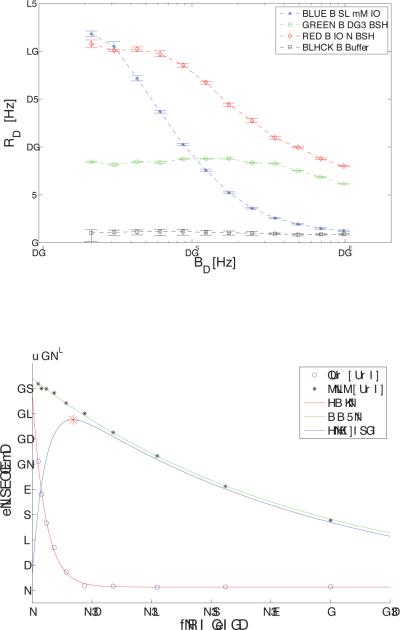
Figure 3.
Shows R1ρ (1/ T1ρ) dispersion differences with ω1 for the X-ray contrast agent Iohexol, BSA, and the combined IO + BSA substance, with the buffer solution for reference.
3.a. 32 mM IO (blue stars), BSA (green circles), IO+BSA (red diamonds), and buffer (black squares) phantoms. Note that the R1ρ dispersion for the IO + BSA phantom was much more marked and occurs at a lower frequency than for BSA alone.
3.b. Time course of signal decay for 32 mM IO phantom at low (250 Hz) and high power (1 kHz). The point of maximum contrast occurs at 137 msec.

