Figure 2.
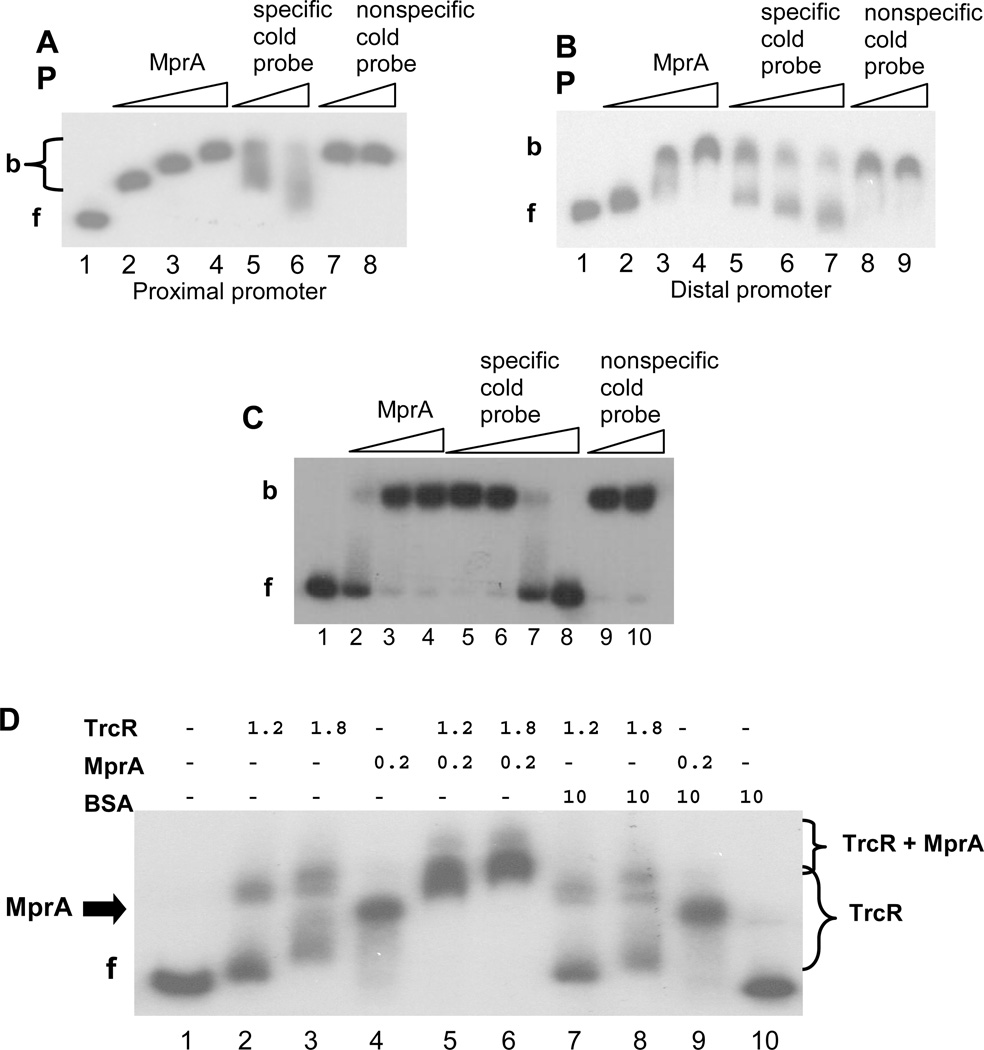
EMSA analyses of MprA binding to the Rv1057 promoter. (A) A 310-bp probe to the proximal region was amplified using primers Rv1057 F1/R4. A fixed amount of probe was incubated with no MprA (Lane 1); 0.25–0.75 µg MprA (Lanes 2–4); 0.75 µg MprA, and 100-fold or 300-fold excess of unlabeled specific probe (Lanes 5 and 6); 0.75 µg MprA, and 100-fold or 200-fold excess of unlabeled nonspecific probe (Lanes 7 and 8). f, free probe; b, bound probe.
(B) A 273-bp probe to the distal region was amplified using primers Rv1057 F12/R10, and a fixed amount was incubated in reaction mixtures containing no MprA (Lane 1); 0.5–1.5 µg MprA (Lanes 2–4); 1.5 µg MprA and 100-fold to 400-fold excess of unlabeled specific probe (Lanes 5–7); 1.5 µg MprA and 50-fold or 200-fold excess of unlabeled nonspecific probe (Lanes 8 and 9).
(C) A 117-bp probe to the proximal region was amplified using primers Rv1057 F4/R5, and a fixed amount was incubated with no MprA (lane 1); 0.5–1.5 µg MprA (Lanes 2–4); 1.5 µg MprA and 100-fold to 400-fold excess of unlabeled specific probe (Lanes 5–8); 1.5 µg MprA and 300-fold or 400-fold of unlabeled nonspecific probe (Lanes 9 and 10).
(D) MprA and TrcR were incubated with the proximal promoter probe F1/R4 separately, or in combination with each other or with BSA. Amounts of each protein are indicated in micrograms. (−) indicates no protein. f, free probe. Brackets indicate range of bands detected in the presence of TrcR alone, or TrcR + MprA, as indicated. The arrow labeled “MprA” marks the position of the band obtained with MprA alone (Lanes 4 and 9).

