Figure 2.
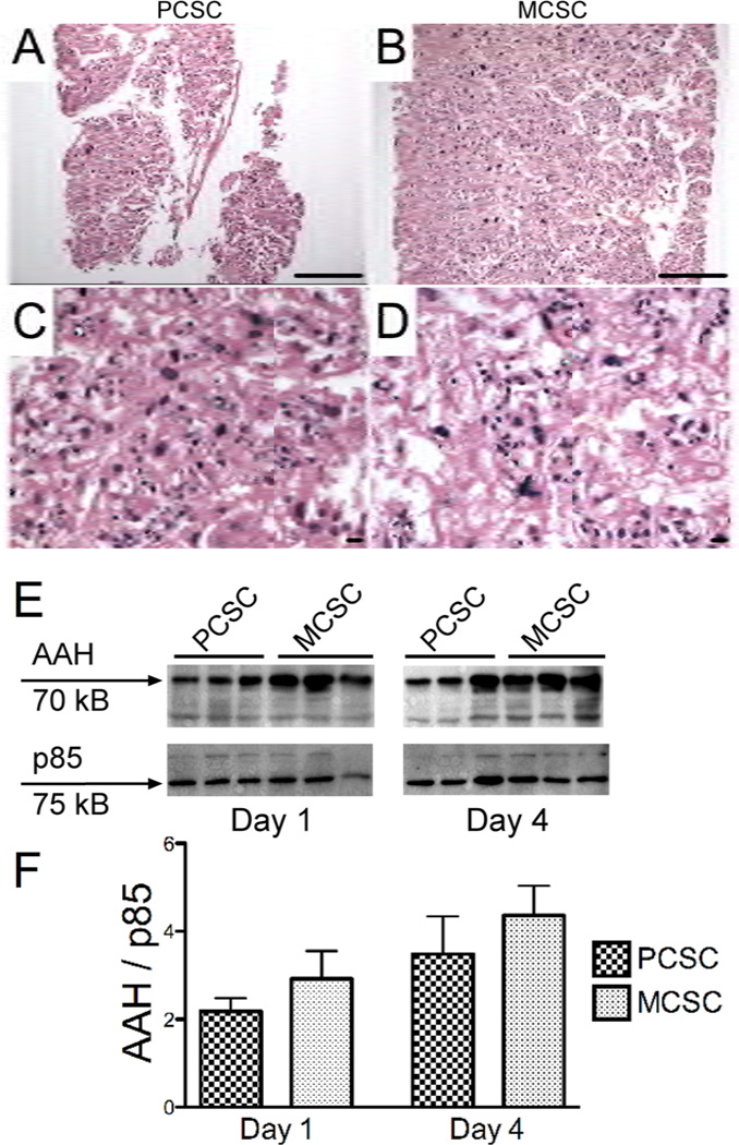
Structural integrity and protein expression in PCSCs and MCSCs. A–D) H&E stained perpendicular plane sections of placental (A) PCSCs and (B) MCSCs demonstrating full-thickness slices of 4-day old cultures. [magnification bar= 25 µm] H&E stained labyrinthine region of (C) PCSCs and (D) MCSCs depicting the central regions of the slices. [magnification bar= 25 µm] E) Western blot analysis of aspartyl-asparaginyl β-hydroxylase (AAH) in placental PCSC and MCSC homogenates harvested after 1 or 4 days in culture. The blots were stripped and re-probed with polyclonal antibody to the p85 subunit of PI3 kinase as a sample loading control. F) Immunoreactivity was quantified by digital imaging and the mean ± S.E.M. AAH/p85 ratios are depicted graphically.

