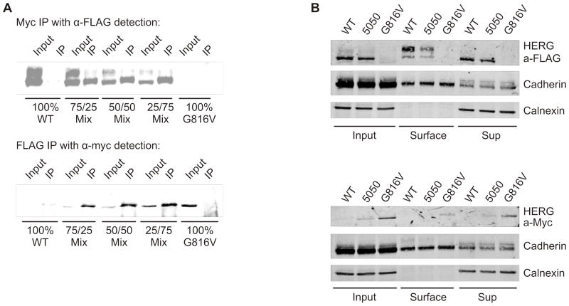
Figure 5. Interaction of WT HERG and G816V HERG subunits.
A) Co-immunoprecipitation and subsequent Western blot from transiently transfected HEK 293 cells shows 3x FLAG-tagged WT HERG detected from a pull-down of myc-tagged G816V HERG in 3 different mixed combinations with 100% WT HERG and 100% G816V HERG as controls, n=3. Lower panel shows the reverse experiment where myc-tagged G816V HERG was detected in a pull-down of 3x FLAG-tagged WT HERG from the same combinations, n=3. B) Immuno-blots of cell surface biotinylation of 3x FLAG-tagged WT HERG and myc-tagged G816V HERG alone or in a 50/50 mix. Proteins were separated by 7.5% SDS-PAGE. Top panel shows a blot with anti-FLAG antibody with cadherin and calnexin as controls. Lower panel shows a blot with anti-Myc antibody with cadherin and calnexin as controls, n=3.

