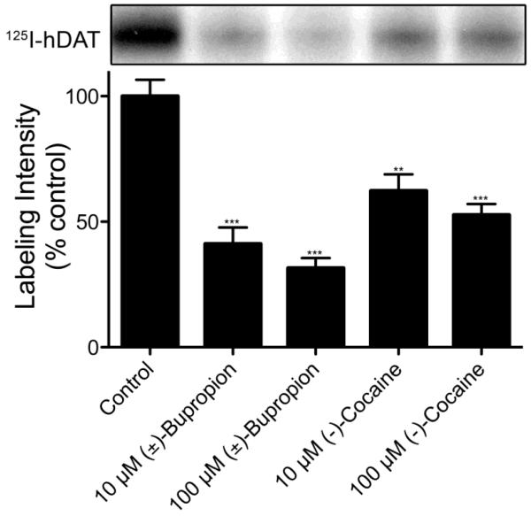
Figure 2.
Photoaffinity labeling of hDAT with (±)-[125I]-3. LLCPK1 cells expressing 6Xhis-hDAT were photoaffinity labeled with 10 nM (±)-[125I]-3 in the absence or presence of 10 μM or 100 μM (±)-bupropion or (−)-cocaine. Cells were solubilized and DATs were immunoprecipitated followed by analysis by SDS-PAGE and autoradiography. The relevant portion of a representative autoradiograph is pictured followed by a histogram that quantitates relative band intensities. (means ± SE of three independent experiments; ***P <0.0001 versus control; **P <0.001 versus control)

