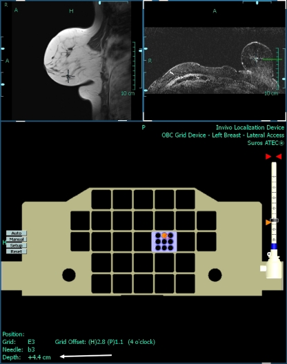
Fig. 2.
Screenshot from a biopsy procedure using a 9 G vacuum-assisted needle to biopsy a small mass-like lesion with an irregular margin at six o’clock in the left breast. The lower screen shows where to put the needle block (purple square) and which hole to use (orange dot) in order to come closest to the optimal position (red circle). The required depth can be read at the bottom of the screen (arrow)

