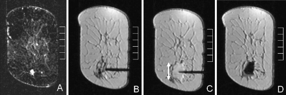
Fig. 6.
Biopsy of a small irregular lesion at 6 o’clock in the left breast using a 9 G vacuum-assisted needle. a subtraction image created before the biopsy procedure to localise the lesion. b placement of the coaxial sheet, the trocar has been removed and replaced with a plastic insert. c the same coaxial sheet directly after the biopsy, note the large haematoma that surrounds the tip of the needle (double-headed arrow). d A large marker is left that also provides some compression from the inside. Histopathological results revealed a complex sclerosing lesion with cystic degeneration. No malignancy was found

