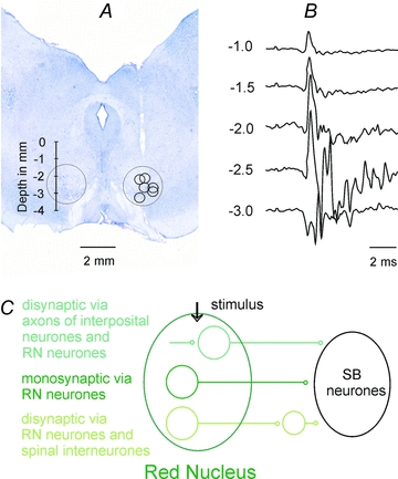
Figure 1. Location of stimulation sites in the contralateral RN.

A, location of electrolytic lesions made at the end of the experiments to mark the stimulation sites overlaid on a transverse section of the midbrain of one of the preparations. The extent of the RN is indicated by the circles, the depths according to Horsley–Clarke's coordinates being indicated to the left.B, a series of antidromic field potentials evoked along the most lateral electrode track to the right recorded at the depths indicated to the left.C, diagram of monosynaptic and two disynaptic pathways via which synaptic actions could be evoked by stimuli applied in RN.
