
Figure 7. Comparison of effects of stimuli applied in the contralateral RN and the contralateral PT before and after the MLF lesion.
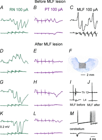
A–C, DandE, GandHandK–M, records from four SB neurones, the first recorded prior the MLF lesion and the three remaining neurones after the lesion. Upper traces, intracellular records from the neurones. Lower traces, records from the cord dorsum.F, the extent of the lesion.IandJ, records from the lateral funiculus at the Th12 and L4 levels showing that the volleys evoked by MLF stimulation disappeared after the lesion. Note that both EPSPs and IPSPs were evoked from the RN after the lesion but only IPSPs followed PT stimuli.