
Figure 7. Small trigeminal ganglion neurons retrogradely labelled from the dura do not include CI-T neurons; most are CS neurons.
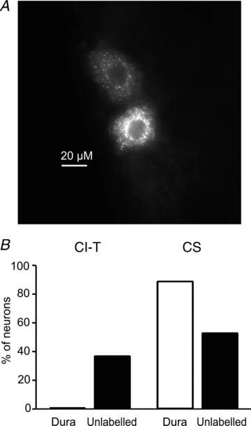
TG neurons were dissociated from wild-type mice in which the fluorescent tracer DiI was previously applied to the dura to label dural afferents. Whole-cell Ca2+ current was recorded from both labelled and unlabelled neurons withC≤ 20 pF. Ca2+ currents were elicited fromVh = –96 mV to test potentials between –66 mV and +54 mV. CI-T neurons were identified on the basis of the presence of LVA Ca2+ current and CS neurons on the basis of capsaicin sensitivity and absence of LVA Ca2+ current.A, image of TG neurons retrogradely labelled from dura from a 16 μm thick slice of a longitudinally sectioned TG. The image was taken using a 40× oil immersion lens.B, fractions of CI-T and CS neurons among small labelled dural and unlabelled TG neurons. The presence of LVA Ca2+ current was tested in 24 labelled dural afferents and 35 unlabelled TG neurons withC≤ 20 pF; 13 of the unlabelled cells were from animals that had surgery but in which DiI was placed in the bone just above the dura. Capsaicin was tested in a fraction of labelled dural (n = 9) and unlabelled (n = 19) TG neurons.