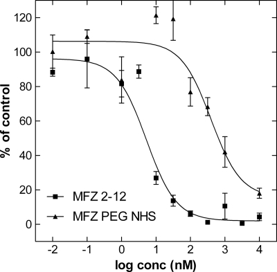
FIGURE 2.
Uptake inhibition experiments comparing MFZ2-12 and a PEGylated form thereof. CHOK1 cells stably expressing SERT were distributed in 48-well plates (5 × 10⁵ cells). The cells were preincubated in Krebs-Ringer-HEPES buffer (0.1 ml) containing either MFZ2-12 or PEG-MFZ2-12 in the concentrations (conc) indicated. After 5 min, [3H]5HT (150 nm) was added, and uptake was allowed for 1 min at room temperature. Uptake was terminated by removing the buffer and washing the cells with ice-cold buffer, and radioactivity was determined by liquid scintillation counting. Data are means ± S.E. of four independent experiments that were carried out in triplicate. Specific uptake in the absence of any inhibitor was 52.1 ± 1.4 pmol·106 cells·min−1 and was set 100% to normalize interassay variations. Nonspecific uptake in the presence of 10 μm paroxetine was <10% of total uptake and subtracted. The solid lines were generated by fitting the data points by non-linear least squares to an equation for the inhibition at a single class of binding sites according to the law of mass action. The estimated IC50 value were 5.26 ± 1.5 nm and 395.8 ± 15.4 nm for MFZ2-12 and its PEG-modified version, respectively.

