FIGURE 3.
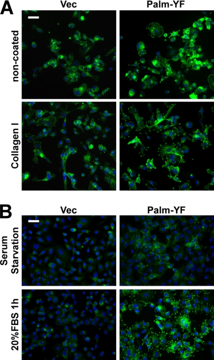
Integrin and growth factor signaling promote formation of peripheral adhesion complexes. A, Palm-PTK6-YF-expressing PC3 cells form more peripheral adhesions on collagen I-coated plates than on non-coated plates. PC3 cells stably expressing Palm-PTK6-YF or vector (Vec) were seeded in collagen I-coated or non-coated chamber slides for 24 h. Cells were stained with anti-phosphotyrosine antibodies (green) and counterstained with DAPI (blue). The size bar denotes 50 μm. B, 1-h 20% FBS stimulation after 24-h serum starvation promotes the formation of peripheral adhesion complexes in Palm-PTK6-YF-expressing PC3 cells. Peripheral adhesion complexes were visualized by anti-phosphotyrosine immunostaining (green). Cells were counterstained with DAPI (blue). The size bar denotes 50 μm.

