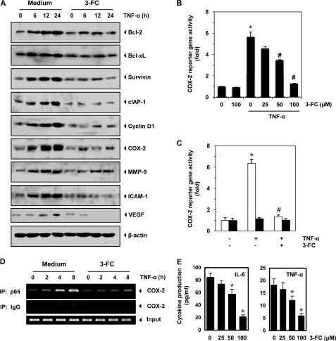
FIGURE 5.
3-FC inhibits expression of TNF-α-induced NF-κB-regulated gene products. A, 3-FC inhibits the expression of TNF-α-induced anti-apoptotic (Bcl-2, Bcl-xL, survivin, and cIAP-1), cell proliferative (cyclin D1 and COX-2), metastatic (MMP-9 and ICAM-1), and angiogenic (VEGF) proteins. KBM-5 cells were incubated with 3-FC (100 μm) for 12 h and then treated with TNF-α for the indicated times. Whole-cell extracts were prepared and analyzed by Western blot analysis using the indicated antibodies. The results shown are representative of three independent experiments. B, 3-FC inhibits the COX-2 promoter activity induced by TNF-α. Cells were transiently transfected with a COX-2 promoter linked to the luciferase reporter gene plasmid for 24 h and then treated with the indicated concentrations of 3-FC for 12 h. Cells were treated with 1 nm TNF-α for an additional 24 h, lysed, and subjected to a luciferase assay. Variations in transfection efficiency were normalized by measuring β-galactosidase activity. The luciferase activity was estimated as luciferase count/β-galactosidase count. The values are the mean ± S.D. for three independent replicates. * and #, significance of difference compared with the control and TNF-α-alone groups, respectively (p < 0.05). C, COX-2 promoter with mutant NF-κB-binding elements is resistant to 3-FC treatment. A293 cells were transfected with a luciferase expression construct ligated to the full-length (white bars) or mutant (black bars) COX-2 promoter. Cells were treated with 3-FC for 12 h, followed by TNF-α for an additional 24 h, and then lysed and subjected to a luciferase assay. The values are the mean ± S.D. for three independent replicates. * and #, significance of difference compared with the control and TNF-α-alone groups, respectively (p < 0.05). D, effect of 3-FC on binding of NF-κB to the COX-2 promoter. Cells were treated with 100 μm 3-FC for 12 h, followed by 1 nm TNF-α for the indicated times, and the proteins were cross-linked with DNA by formaldehyde and subjected to ChIP assay using anti-p65 antibody and the COX-2 primer. Reaction products were resolved by electrophoresis. IP, immunoprecipitate. E, 3-FC down-regulates IL-6 and TNF-α production in U266 cells. Cells were treated with the indicated concentrations of 3-FC, and cell free supernatants were harvested after 12 h. The levels of IL-6 and TNF-α were detected by ELISA. The values are the mean ± S.D. for three independent replicates. *, significance of difference compared with the control (p < 0.05).

