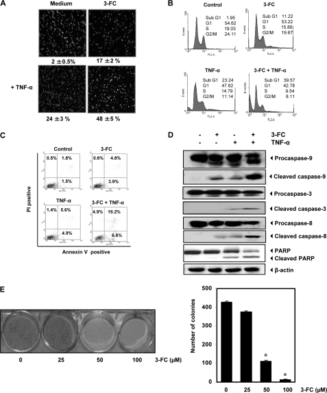
FIGURE 6.
3-FC potentiates apoptotic effects of TNF-α. A–C, KBM-5 cells were pretreated with 3-FC for 12 h and then with TNF-α (1 nm) for 24 h. Cell death was determined by LIVE/DEAD assay (A), sub-G1 analysis (B), and PS externalization assay (C). Values below each photomicrograph in A represent the mean ± S.D. of apoptotic cells. PI, propidium iodide. D, 3-FC potentiates TNF-α-induced caspase activation and PARP cleavage. KBM-5 cells were incubated with 50 μm 3-FC for 12 h and then treated with 1 nm TNF-α for 24 h. Whole-cell extracts were prepared and analyzed by Western blotting with the indicated antibodies. E, 3-FC suppresses long-term colony formation by tumor cells. Cells were treated with 3-FC for 12 h, washed, trypsinized, and reseeded in 100-mm dishes. After 14 days, colonies were stained with crystal violet and counted. The values are the mean ± S.D. for three independent replicates. *, significance of difference compared with the control (p < 0.05).

