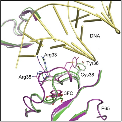
FIGURE 7.
Possible binding mode of 3-FC with p65 DNA-binding region. The original crystal structure (Protein Data Bank code 1VKX) is superimposed with the modeled structures of the wild-type and C38S mutant proteins upon 3-FC binding. Gray, original crystal structure; purple, modeled wild-type structure upon 3-FC binding; green, modeled C38S mutant upon 3-FC binding. The final docked pose for 3-FC is depicted in purple and green sticks for the wild-type and C38S mutant structures, respectively. DNA is represented as yellow tubes.

