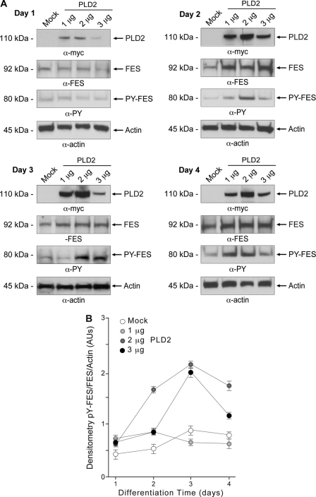
FIGURE 6.
PLD2 overexpression in differentiating HL-60 cells increases Fes expression and phosphorylation. HL-60 cells were nucleofected with constructs encoding for PLD2-WT or mock-transfected and immediately induced to differentiate in fresh media with 1.25% DMSO. A, protein expression and phosphorylation were ascertained in protein extracts obtained at days 1–4. Protein samples were subjected to SDS-PAGE and electroblotted. Membranes were developed by using antibodies directed against PLD2, Fes, or phosphorylated Fes on tyrosine residues (PY-Fes). To ensure equal loading, the same membranes were probed against α-actin. Sample size for each experiment was n = 3. B, densitometric analysis of Fes phosphorylation. The densitometric ratios for phospho-Fes/Fes/β-actin are plotted. Data are means ± S.E. AUs, arbitrary units.

