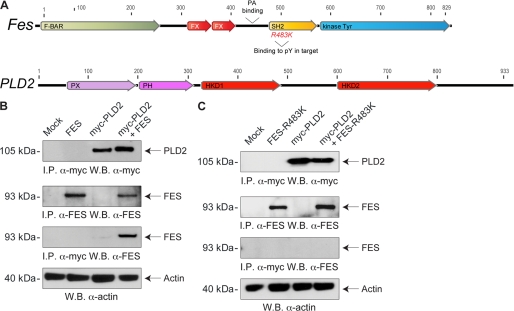
FIGURE 9.
PLD2 interacts with Fes in vivo. A, schematic drawings of human Fes (NP_001996, top) and PLD2 (NP_002654, bottom) proteins to show the predicted motifs (F-BAR and FX (a module that recognizes positive membrane curvature), SH2 (Src homology domain 2 recognizes phosphotyrosine residues with an invariant arginine) and kinase Tyr (the putative tyrosine kinase domain)). PLD2 protein drawing shows its classic domains, PH (pleckstrin homology domain), PX (phosphoinositide-binding domain, p40phox/p47phox-homology domain), and HKD1 and HKD2 (the two catalytic sites of PLD2). B and C, cells were transiently transfected with plasmids encoding for Myc-tagged PLD2 (myc-PLD2), human Fes (WT (B) or SH2-compromised R483K (C)) alone or in combination. Two days post-transfection, protein lysates were obtained and immunoprecipitated (I.P.) with anti-human Fes or anti-Myc antibodies. Immunoprecipitates were separated by SDS-PAGE and immunoblotted with anti-Myc antibodies or anti-Fes, respectively. W.B., Western blot.

