FIGURE 2.
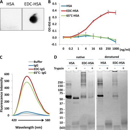
Characterization of EDC-crosslinked proteins. A, membrane-bound native HSA and EDC-crosslinked HSA were examined with anti-Aβ-oligomer (A11) antibody by dot blot analysis. B, direct binding of A11 antibody to different amounts of native, EDC-crosslinked, or 65 °C-aggregated HSA pre-coated on an ELISA plate. Error bars are means ± S.D. of absorbance at 450 nm. C, fluorescence emission profiles of bis-ANS obtained after incubating with various IgG-derived proteins in comparison with PBS buffer. D, SDS-PAGE separation of HSA and EDC-crosslinked HSA, which had been subjected to partial trypsin digestion for 60 min (Native, left). As a control, both proteins were denatured by boiling prior to the identical trypsin digestion procedure (Denatured, right).

