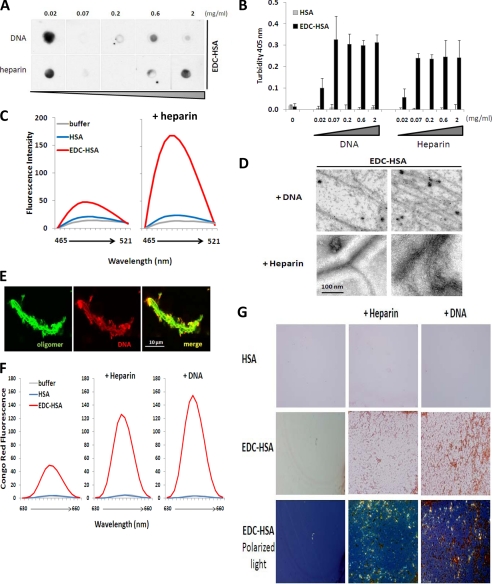
FIGURE 4.
Binding with nucleic acids and heparin converts EDC-crosslinked proteins to amyloid. EDC-crosslinked HSA (0.2 mg/ml) was incubated with different amounts of DNA or heparin. The resulting samples were analyzed by (A) dot blot with A11 antibody and (B) OD at 405 nm to measure the solution turbidity. C, native or EDC-crosslinked HSA were incubated with buffer or heparin, before mixing with dye ThT. Differential fluorescence emission profiles of the resulting samples are shown. D, transmission electron microscopy analysis of EDC-crosslinked HSA complexed with either DNA (top) or heparin (bottom). E, confocal imaging analysis of EDC-crosslinked IgG (green) complexed with fluorescently labeled DNA (red). F, and G, native or EDC-crosslinked HSA were incubated with buffer, heparin, or DNA, before mixing with dye Congo Red. Differential fluorescence emission profiles (F) and microscopic analysis (G) of the resulting samples are shown. In G top two rows: brightfield light; bottom row: transmitted polarized light (bottom row). Magnification, ×40.

