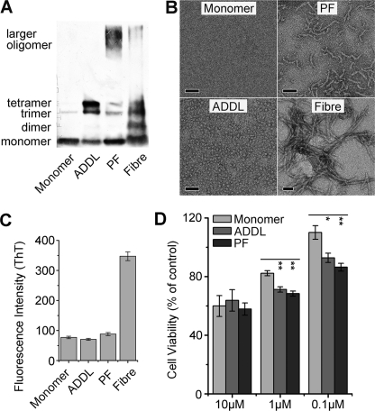
FIGURE 1.
Characterization of Aβ samples in different assembly states. The characteristics of Aβ monomer, ADDL, protofibril (PF), and fiber were assessed by immunoblotting with 6E10 (A), EM observation (scale bars represent 50 nm) (B), and thioflavin T (ThT) fluorescence (C). Aβ monomer primarily migrated as a single band at ∼4.5 kDa in SDS-PAGE and was invisible under EM, whereas ADDL was characterized by bands corresponding to trimer/tetramer in SDS-PAGE and particle-like appearances with even size distribution (∼5 nm) under EM. There were additional high molecular weight bands in PF and fiber, and these two samples showed expected morphology. The absence of fiber in Aβ monomer, ADDL, and PF preparations were further verified by the comparable low ThT fluorescence as compared with that of mature fiber. D, cell viability of N2a cells treated with different sterile Aβ species for 48 h was assessed by 3-(4,5-dimethylthiazol-2-yl)-2,5-diphenyltetrazolium bromide assay. Results (n > 3) are given as mean ± S.E.; *, p < 0.05; **, p < 0.005. At low concentrations PF and ADDL are significantly more toxic than monomer sample in cell viability assay.

