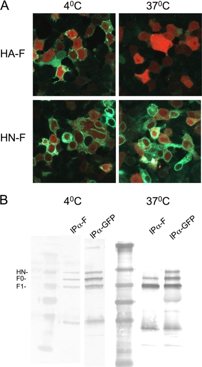
FIGURE 7.
Temperature dependence of BiFC and co-immunoprecipitation. 293T cells were co-transfected with constructs encoding either HN wt N-Venus and F C-CFP or HA N-Venus and F C-CFP and treated overnight with 10 mm zanamivir and before analysis fresh 10 mm zanamivir and cycloheximide were added. A, the cells were incubated for 1 h at either 23 °C or 37 °C to allow protein maturation and then transferred to either 4 or 37 °C. Green fluorescence indicates BiFC for HA-F at 4 °C, but not at 37 °C, and BiFC for HN-F at both temperatures. B, the cells were lysed at either 4 or 37 °C. The figure shows the Western blot after immunoprecipitation (IP) with either anti-F (to show coimmunoprecipitation of the BiFC partners) antibodies or anti-GFP antibodies (control for expression and immunoprecipitation).

