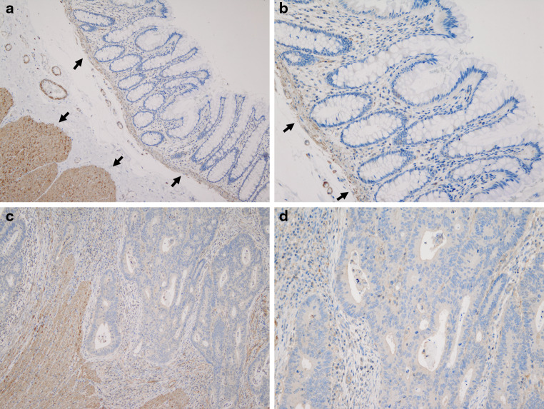
Fig. 1.
Immunohistochemistry of TLR4 in NCT (×10, a) (×20, b) and CT (×10, c) (×20, d). All layers of the tissue specimens were stained, and TLR4 was strongly detected in the proper muscular layer and the proper mucosal layer (arrows) (a, c). TLR4 was less positively stained in the lamina propria of the mucosa in both NCT and CT (b, d)

