Fig. 3.

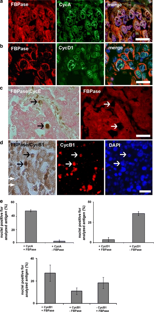
Nuclear localization of FBPase in different phases of the cell cycle in human squamous cell lung cancer. a Co-localization of CycA and FBPase in nuclei of S/G2 phase cells. b Localization of FBPase and G1 phase marker, CycD1. c Co-immunostaining for FBPase and CycE, a marker of G1/S transition. Arrows indicate CycE-positive and FBPase-negative cells. d Co-localization of CycB1 and FBPase. Arrows point to FBPase- and CycB1-positive nuclei. e Percentage of nuclei positive for CycA, CycD1 and CycB1 with respect to subcellular localization of FBPase. In a, b and d the nuclei were counterstained with DAPI. Bar 30 μm in a, b and 40 μm in c, d
