
Fig. 5.
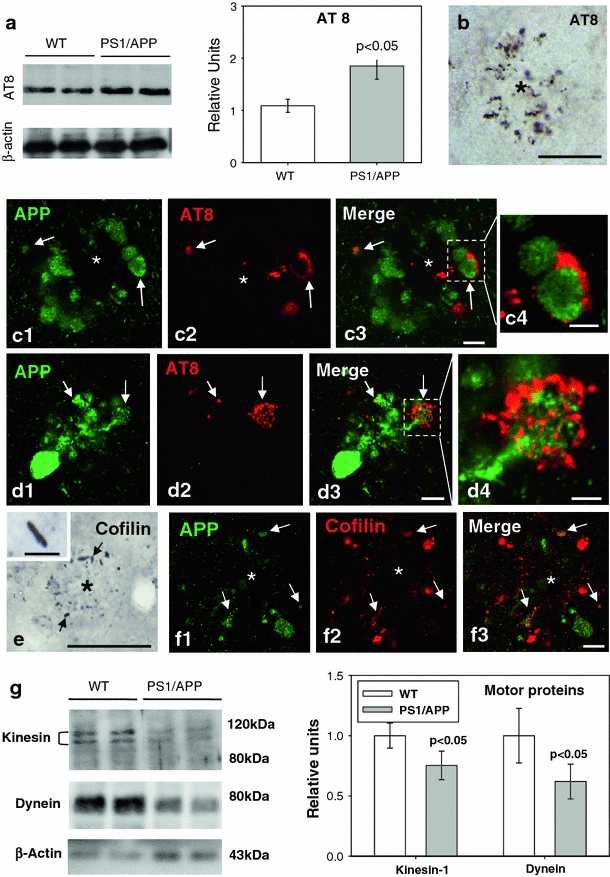
Cytoskeletal pathology in young PS1/APP hippocampus. a Quantitative Western blotting using AT8 antibody in the hippocampus of 6-month-old PS1/APP and WT mice; PS1/APP mice had significantly higher AT8 levels than to age-matched WT mice (n = 6, p < 0.05). b The presence of AT8-positive structures around plaques (asterisk) was observed from 4 months of age. c1–c4 and d1–d4. Double-label immunofluorescence confocal laser microscopy of APP (green) and AT8 (red) in 6-month-old transgenic mice showing the presence of phospho-tau in some APP-positive dystrophic neurites. e Immunohistochemistry for cofilin revealed the presence of actin–cofilin rods around plaques (asterisk) from 4 months of age. A higher magnification detail of a rod is shown in the inset. f1–f3 Confocal laser microscopy with double APP/cofilin labeling revealed the presence of cofilin within some APP-positive neurites. g Quantitative Western blotting for heavy chain kinesin-1 and dynein in 6-month-old PS1/APP and WT mice hippocampus. PS1/APP mice were found to have significantly lower kinesin-1 and dynein levels than age-matched WT mice (n = 8, p < 0.05). Scale bars b 100 μm, c1–c3 5 μm, c4 2.5 μm, d1–d3 7.5 μm, d4 2.5 μm, e 50 μm (inset 7 μm), f1–f3 7.5 μm