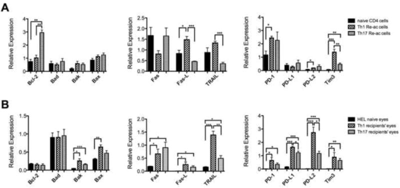
Fig. 2.

Expression profiles of apoptosis-related molecules are different between reactivated Th1 and Th17 cells in culture and between mouse eyes with inflammation induced by Th1 or Th17 cells. Levels of apoptosis-related molecule transcripts were measured by qPCR in cultures of activated/polarized Th1 or Th17 cells re-exposed to HEL (A), or eyes of recipient mice with inflammation induced by adoptive transfer of reactivated Th1 or Th17 cells (B). Cultures of naïve CD4 cells, with no activation, were used as controls in the experiments with cultured cells (A), whereas eyes of naïve control mice served as controls in the experiments with inflamed eyes, summarized in B. The columns are means ± SEM of four experiments each.
