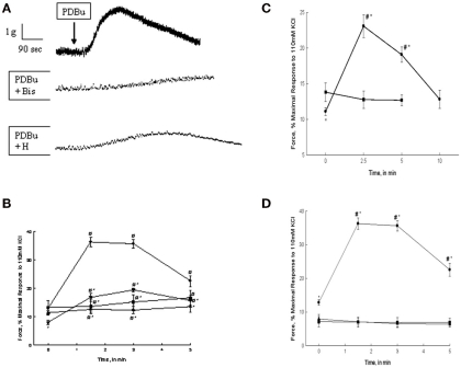
Figure 1.
PDBu, α-PDBu, or DOG-stimulated bladder smooth muscle contraction and the effects of PKC or ROCK inhibition or calcium removal. (A) Representative tracing of contractions in response to 3 μM PDBu alone (top tracing), and in the presence of 3 μM Bis (middle tracing), or 1 μM H-1152. Arrow denotes the addition of PDBu, inhibitors were added 20 min prior to stimulation. (B) Intact strips of bladder smooth muscle were contracted for 5 min with PDBu (3 μM) in the presence or absence of Bis (3 μM) or H-1152 (1 μM). Strips contracted in response to PDBu alone (•) generated 36.5 + 2.2% of maximal force at ∼1.5 min and maintained a sustained contraction until ∼3 min. Inhibition with Bis (▲) or H-1152 (▼) significantly decreased force at 1.5 and 3 min. H-1152 also significantly decreased basal force at 0 min. Stimulation with α-PDBu (■) did not elicit an increase in force until 5 min. (C) Intact strips of bladder smooth muscle were contracted for 10 min with DOG (300 μM). DOG (•) generated force significantly greater than basal values at 2.5 and 5 min of stimulation, but not significantly different from basal values at 10 min. Vehicle alone (DMSO, ■) had no effect. (D) Intact strips of bladder smooth muscle were contracted for 5 min with PDBu (3 μM, •) in the presence or absence of extracellular calcium or after depletion of all calcium stores. Strips contracted in the absence of extracellular calcium (■) or following the depletion of all calcium stores (▲) did not respond to PDBu. Values shown are the means ± SE of at least five determinations. *P < 0.05, compared with values in the presence of PDBu alone (B), DMSO alone (C), or the absence of calcium (D). #P < 0.05, compared with values at 0 min of the same treatment.

