
Fig. 2.
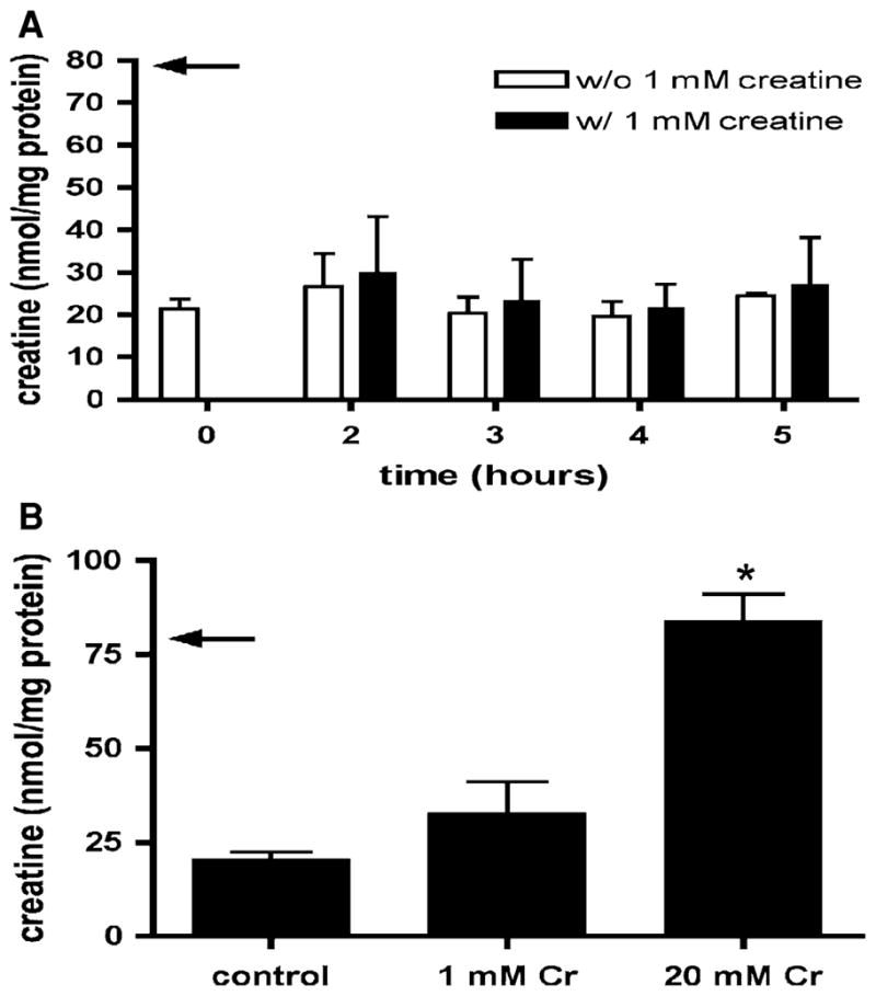
a Cr content of isolated mouse cardiomyocytes during incubation in culture medium with and without 1 mM Cr for up to 5 h. The arrow indicates the value for isolated perfused heart tissue (from [34]). Over the time frame of this experiment isolated cardiomyocytes were unable to increase their intracellular Cr. Means ± SE from three independent experiments. b Cr content of isolated cardiomyocytes from un-supplemented isolation buffers, and buffers supplemented with Cr 1 and Cr 20. Addition of 20 mM Cr is sufficient to maintain normal Cr levels (arrow indicates value for isolated perfused heart tissue (from [34])). Means ± SE from 5 to 7 independent myocyte preparations. *P < 0.001 versus without Cr and 1 mM Cr, one-way ANOVA