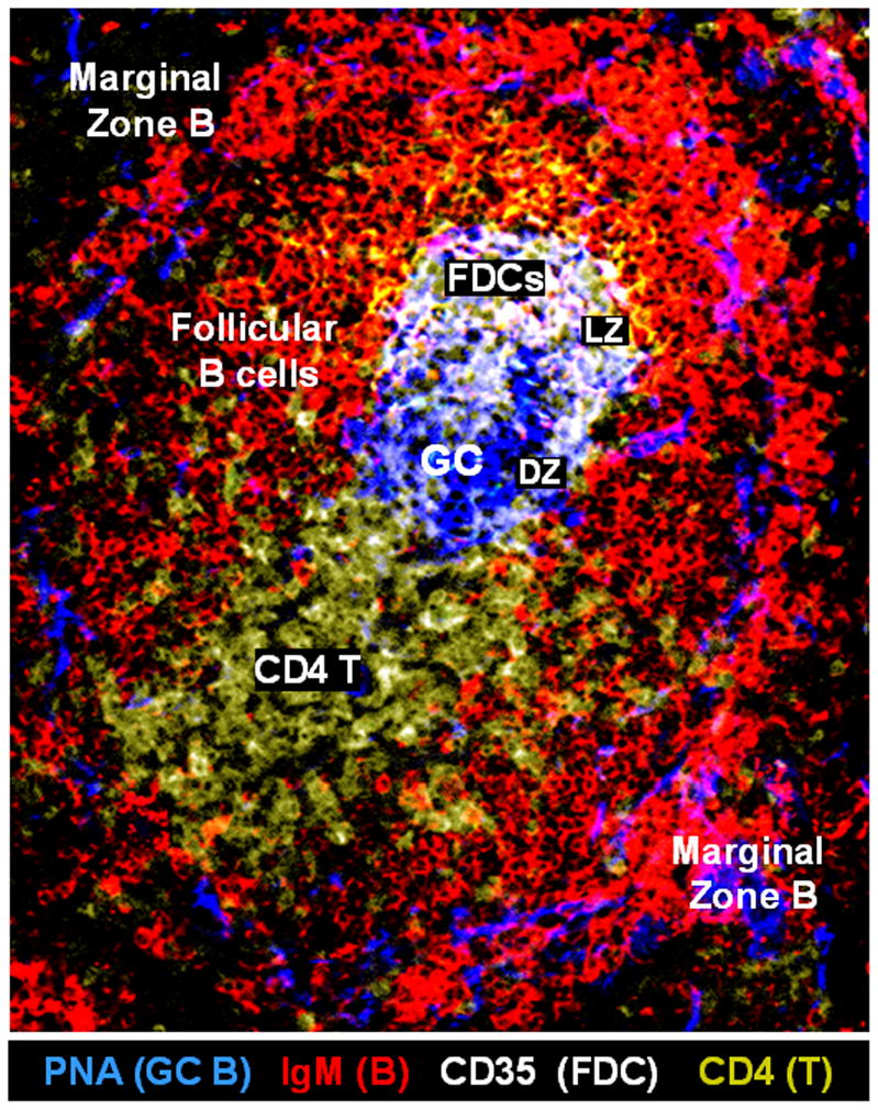
Figure 1.

Anatomic structure of a spontaneous germinal center (GC) in the spleen of a representative BXD2 mouse. Confocal image of a representative BXD2 spleen section stained with anti-IgM (red), anti-CD35 (white), and anti-CD4 (yellow) antibodies, and peanut agglutinin (PNA) (blue). Anti-CD35 antibodies are used to stain follicular dendritic cells (FDCs) that express CD35 (DZ: dark zone; FDCs: follicular dendritic cells; GC: germinal center; LZ: light zone; PNA: peanut agglutinin).
