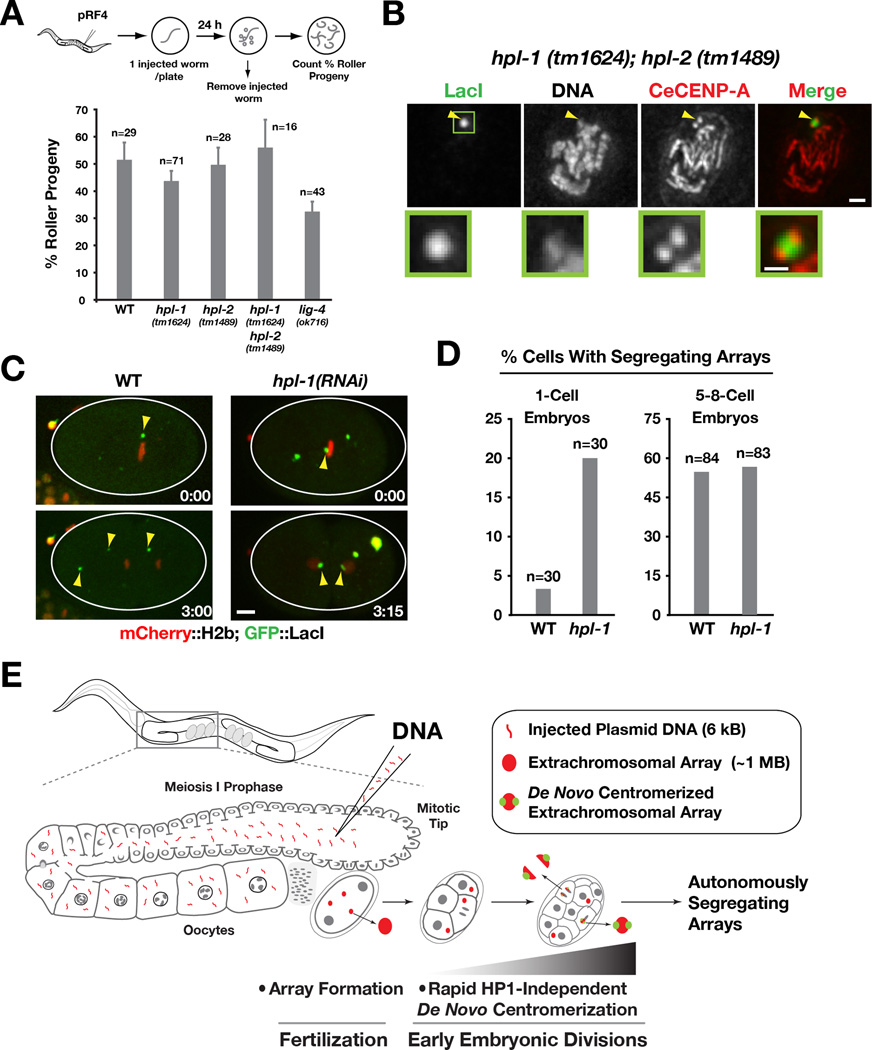
Figure 4. Heterochromatin protein 1 (HPL-1/HPL-2) mutants do not affect array formation or de novo centromerization.
A) Bar graph showing the percentage of Roller progeny produced during the first 24 hrs post-injection of pRF4 in wild-type, hpl-1(tm1624), hpl-2(tm1489), hpl-1(tm1624);hpl-2(tm1489) and lig-4(ok716) mutants. The number of worms analyzed in each condition (n) is indicated. Error bar represents 95% confidence interval for the mean.
B) CeCENP-A localizes to opposing faces of LacO-containing extrachromosomal arrays that have been generated and propagated in the hpl-1(tm1624);hpl-2(tm1489) double mutant. Arrowheads point to the array; the boxed region is magnified below. Scale bar 1 µm (0.5 µm for magnified regions).
C) Examples of 1-cell embryos in wild-type and hpl-1(RNAi), dissected and imaged 4–8 h after p64xLacO injection. hpl-1 RNAi was performed as in Fig. 3B, except that worms were recovered for 20h prior to p64xLacO injection. The wild-type embryo image is the same as in Fig. 2B. Scale bar 5 µm.
D) Bar graph showing the percentage of cells with a segregating array in wild-type and hpl-1 inhibited (using either the mutant or RNAi) at the1-cell stage and at the 5 to 8-cell stage. The number of cells analyzed at each stage (n) is indicated. Note that the Y-axis is modified to facilitate comparison of the two conditions at the two different embryo stages.
E) Model summarizing the key findings. Array formation occurs immediately after fertilization but array centromerization occurs over a longer time scale. HP1 family proteins are dispensable for both array formation and centromerization.

