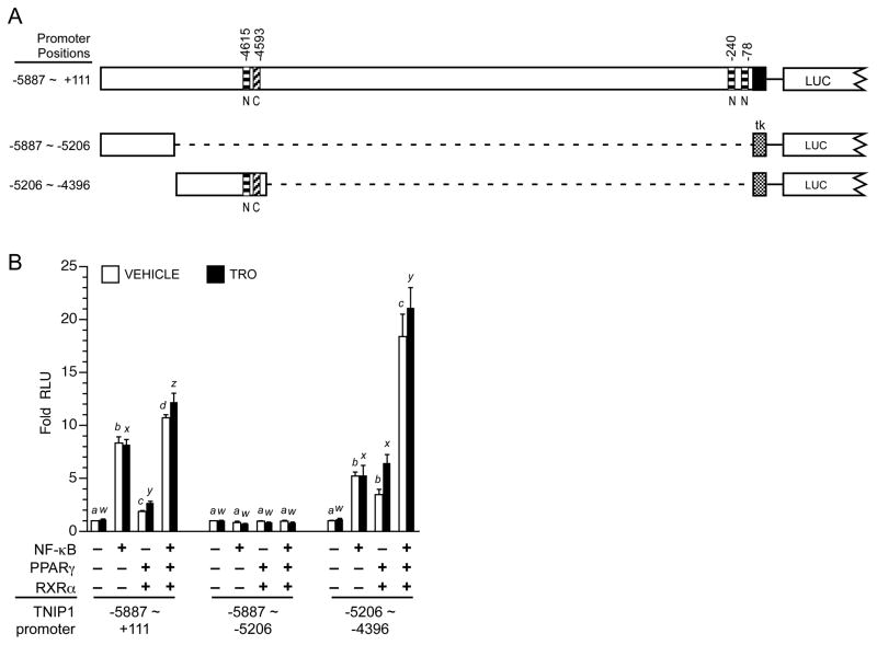
Fig. 8. Individually and together, NF-κB and PPARγ stimulate TNIP1 promoter reporter activity.
(A) Schematic of TNIP1 promoter constructs. Diagonal striped bar, element C PPRE. Horizontal striped bars, NF-κB. Stippled box, tk minimal promoter. LUC, 5′ end of luciferase gene. Distal promoter fragments were ligated (dashed line) to tk but are shown aligned to their native position in the 6kb construct for ease of comparison.
(B) Combined NF-κB and PPARγ activation of TNIP1 promoter. HeLa cells were transfected with equal copy number of the indicated TNIP1 promoter constructs. The two distal fragments utilized the tk minimal promoter (see panel A). Cells were cotransfected with empty expression vector as control (first bar pair, each set) or constructs expressing NF-κB (p50 and p65), or PPARγ and RXRα, or both. Normalized RLU from each TNIP1 promoter construct for control empty expression vector transfection, vehicle-treated media culture condition, was set at one for each reporter. Change in expression for each reporter is expressed as fold of its respective empty vector, vehicle control. Error bars are SEM. Open bars: vehicle control. Solid bars: PPARγ ligand, 1μM troglitazone (TRO). For statistical tests, results were compared within reporter constructs and either within vehicle (a, b, etc) or ligand treatments (w, x, etc). Different letters indicate different values, p<0.05, ANOVA, Newman-Keuls multiple comparison test.

