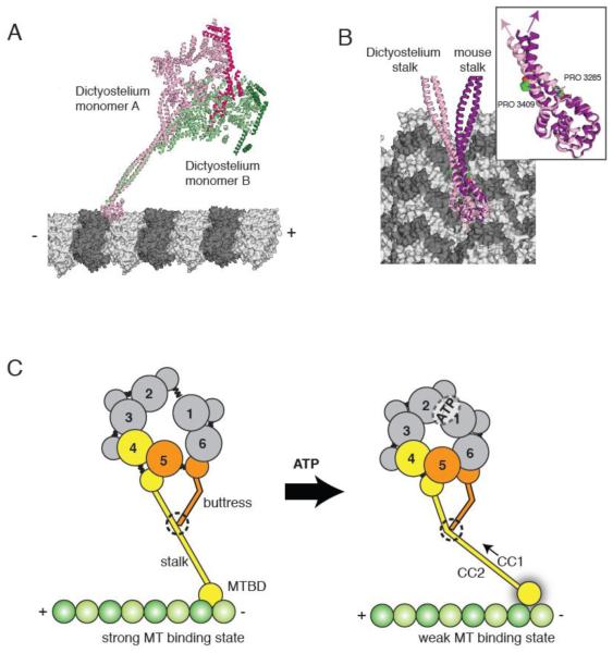
Figure 5.
(A) The two Dictyostelium dynein monomers (monomer A in pink monomer B in green; linkers highlighted in darker hues) modeled onto microtubules based on docking of the mouse microtubule binding domain as in [30]. The different stalk angles of monomer A and B results in a different orientation of the dynein heads with respect to the microtubule longitudinal axis. (B) The stalks of the Dictyostelium stalk (pink) and mouse stalk (purple) viewed from the microtubule minus end. The inset shows the difference in stalk angle, which might be due to differences in the vicinity of the proline kinks in CC1 and CC2. (C) A model for stalk communication. Upon ATP binding, movements of AAA4 and AAA5 small domains are relayed to the stalk and buttress coiled coil extensions, respectively. Due to its interaction between the stalk, the buttress can push or pull on CC1, causing relative sliding motions between CC1 and CC2 and ultimately changing the microtubule binding affinity at the tip of the stalk.

