
Fig. 1.
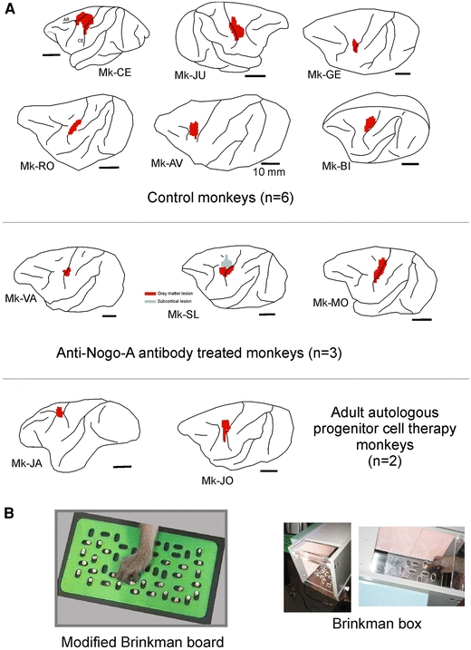
a Location and extent of the permanent unilateral lesion of the M1 hand representation as seen on corresponding lateral views of the brain for 11 monkeys included in the present study (see Table 1). The lesion territory is represented in red, as derived from the lesioned zone of cerebral cortex (gray matter) visible on consecutive frontal histological sections. Spread of the lesion to the subcortical white matter below the gray matter is not represented here, except in monkey Mk-SL in which a subcortical white matter territory was lesioned (gray spot), in a region located more medially than the red territory. The motor cortex lesion was performed in all monkeys on the left hemisphere, except in Mk-JU in which the lesion was in the right hemisphere. Six monkeys (top panel) were control animals for two pilot treatment studies: three monkeys were treated with anti-Nogo-A antibody (middle panel) whereas two monkeys were subjected to an autologous cell therapy (see “Methods” and Table 1). b View of the Modified Brinkman Board (left) and the Brinkman box (right)