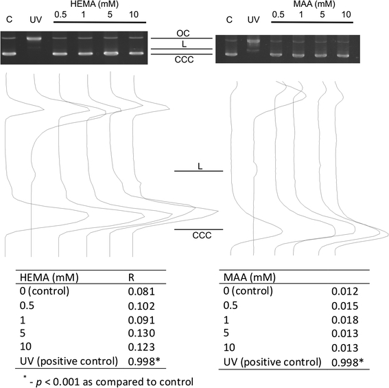
Fig. 3.

DNA damage in isolated pUC19 plasmid. The picture shows three forms of the plasmid: linear (L), open circular (OC) and supercoiled (CCC) exposed to 2-hydroxyethyl methacrylate (HEMA) or methacrylic acid (MAA) at indicated concentrations or 254 nm UV at the dose rate of 0.12 J m−2 s−1 (positive control). The samples were electrophoresed on a 1% agarose gel, stained with ethidium bromide and visualized in UV light. The tables display the ratio of the open circular DNA to the total amount of DNA (R) of the isolated pUC19 plasmid exposed to HEMA, MAA or UV. The values for supercoiled DNA were multiplied by 1.66 to correct for the decreased intercalating ability of ethidium bromide
