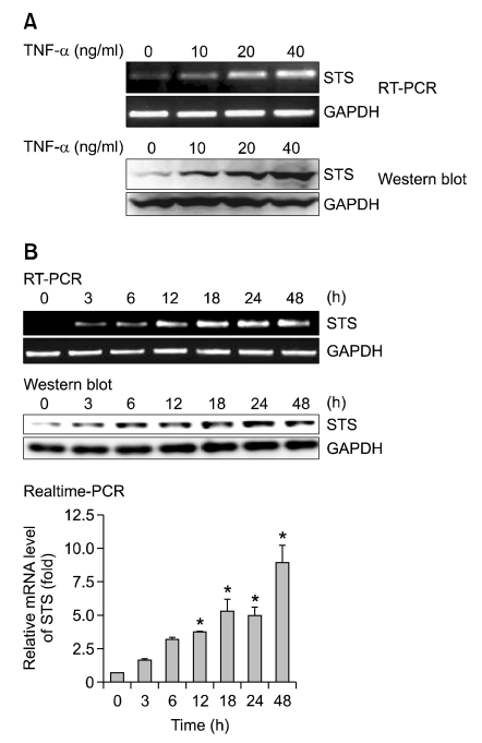
Figure 1.
TNF-α induces STS mRNA and protein expression in PC-3 cells. (A) PC-3 cells were treated with various concentrations of TNF-α (0, 10, 20 or 40 ng/ml) for 18 h. Total RNA was isolated and expression of STS mRNA was assessed using RT-PCR. Expression of GAPDH mRNA was determined as RNA control. For Western blot, total cellular lysates were prepared and STS protein expression was assessed by STS antibody. GAPDH was used as a loading control. (B) Cells were incubated at 37℃ with TNF-α (40 ng/ml) for various time intervals. Expression of STS mRNA was assessed using RT-PCR and real-time PCR analysis. The data shown represent the fold increase in the levels of STS mRNA relative to the control after normalization with GAPDH mRNA level. *P < 0.05 compared with the control cells. The protein levels of STS were determined by Western blot analysis.

