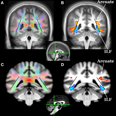
Figure 2.
Location of arcuate (yellow–orange) and inferior longitudinal fasciculi (ILF, blue) in (A,B) humans and (C,D) chimpanzees as revealed by diffusion tractography. Coronal sections for each species are at the posterior aspect of the splenium (see mid-sagittal insets). Tracts include voxels in which 33% or more subjects have a pathway above threshold (0.1% of waytotal). The black lines indicate the angle of the ILF in humans and chimpanzees. The white dotted line in (A) shows the angle of the ILF of chimps overlaid on the human color FA map. In humans, the arcuate appears to have displaced the ILF in a ventromedial direction.

