
Figure 5.
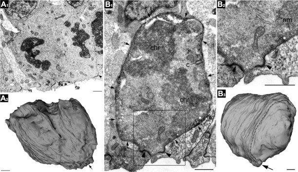
3D reconstructions of SNPs by EM. A1, A2, This short cell in telophase was reconstructed from serial sections taken from unstained tissue from an E13.5 neocortex. The membrane of the cell is indicated with small arrows. The ventricular surface is denoted by a large arrow in the surface rendered image in A2. Adherens junctions are indicated by arrowheads. B1, This E13.5 VZ cell was first labeled by in utero electroporation with EGFP-F. Subsequent anti-EGFP immunolabeling demarcated the cell border (small arrows) with electron dense immunoperoxidase-DAB reaction end-product. Two centrioles (c) located in controversial poles of the cell body (only one of which is seen in this serial section), chromosomes (chr) in cytoplasm, and fragments of forming nuclear membranes (nm) indicate that the cell is in early telophase. The framed area in B1 is enlarged in B2. B3, 3D reconstruction from 144 contiguous serial sections demonstrates that the cell is devoid of processes. The ultrathin section in B1 and B2 is indicated by the dashed line in B3. Scale bars: A1–B3, 1 μm.