
Figure 8.
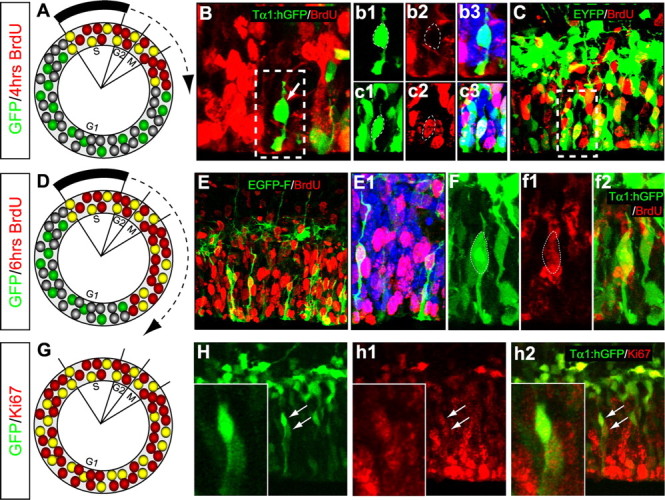
SNPs progress through the cell cycle. The denoted plasmid constructs were used for in utero electroporation on E14.5 followed 24 h later by cumulative labeling with BrdU or immunostaining for Ki67 antigen. A–C, Four hours of BrdU labeling identified bipolar SNPs that were labeled or unlabeled with BrdU. As depicted in A, BrdU+ cells would be present in phases S-early G1 based on cell cycle parameters specified by Takahashi et al. (1995). Correspondingly, BrdU– short cells would be in mid to late G1 phase. B, b1–b3, This Tα1:hGFP expressing cell was BrdU– and therefore in G1 phase, whereas the pEYFP-C2 expressing cell in C and c1–c3 was BrdU+. D, Depiction of the cell-cycle location of BrdU+ and BrdU– VZ cells after 6 h labeling. E–f2, Examples of BrdU+ SNPs reconstructed after transfection with pEGFP-F and Tα1:hGFP. More SNPs were BrdU+ after 6 h of cumulative BrdU exposure compared with 4 h BrdU exposure, suggesting that SNPs progress through the cell cycle (see text for details). G–h2, Ki67 immunostaining also revealed that Tα1-expressing VZ cells are arrayed throughout the cell cycle. The higher magnification insets in H–H2 demonstrate two Tα1-expressing cells with abventricular somata that are Ki67+.