Figure.
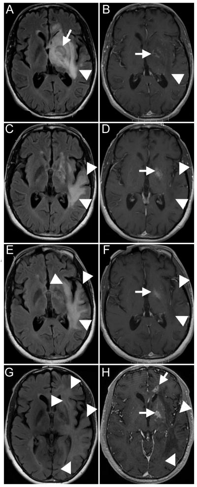
(A–B) MRI at the time of initial presentation. Fluid Attenuated Inverse Recovery image (FLAIR) (A) shows a toxoplasmosis lesion in the left basal ganglia (arrow), with surrounding hyperintense signal, attributed to edema (arrowhead). Post-gadolinium T1 image (B) shows ring enhancement around the toxoplasmosis lesion (arrow), and hypointense signal in the surrounding area (arrowhead). (C–D) MRI obtained one month after original presentation. FLAIR sequence (C) shows almost complete resolution of the left basal ganglia lesion (arrow), but progression of the white matter hyperintensities (arrowheads). Post-biopsy changes are seen in the left basal ganglia on T1 with gadolinium (D, arrow), which were also present on pre-contrast T1 images (not shown). There is no enhancement of the temporal lobe lesions (D, arrowheads). (E–F) Third MRI shows further progression of the hyperintense lesions on FLAIR (E, arrowheads), which where hypointense and did not enhance with contrast on T1 images (F, arrowheads). The original toxoplasmosis lesion is indicated by the arrow in F. (G–H) Last MRI shows again progression of the white matter lesions of PML on FLAIR (G, arrowheads), which remain hypointense on magnetization-prepared 180 degrees radio-frequency pulses and rapid gradient-echo T1 sequence (H, arrowheads). There is new contrast enhancement in the basal ganglia and in the anterior corpus callosum (H, arrows), suggestive of a recurrence of toxoplasmosis.

