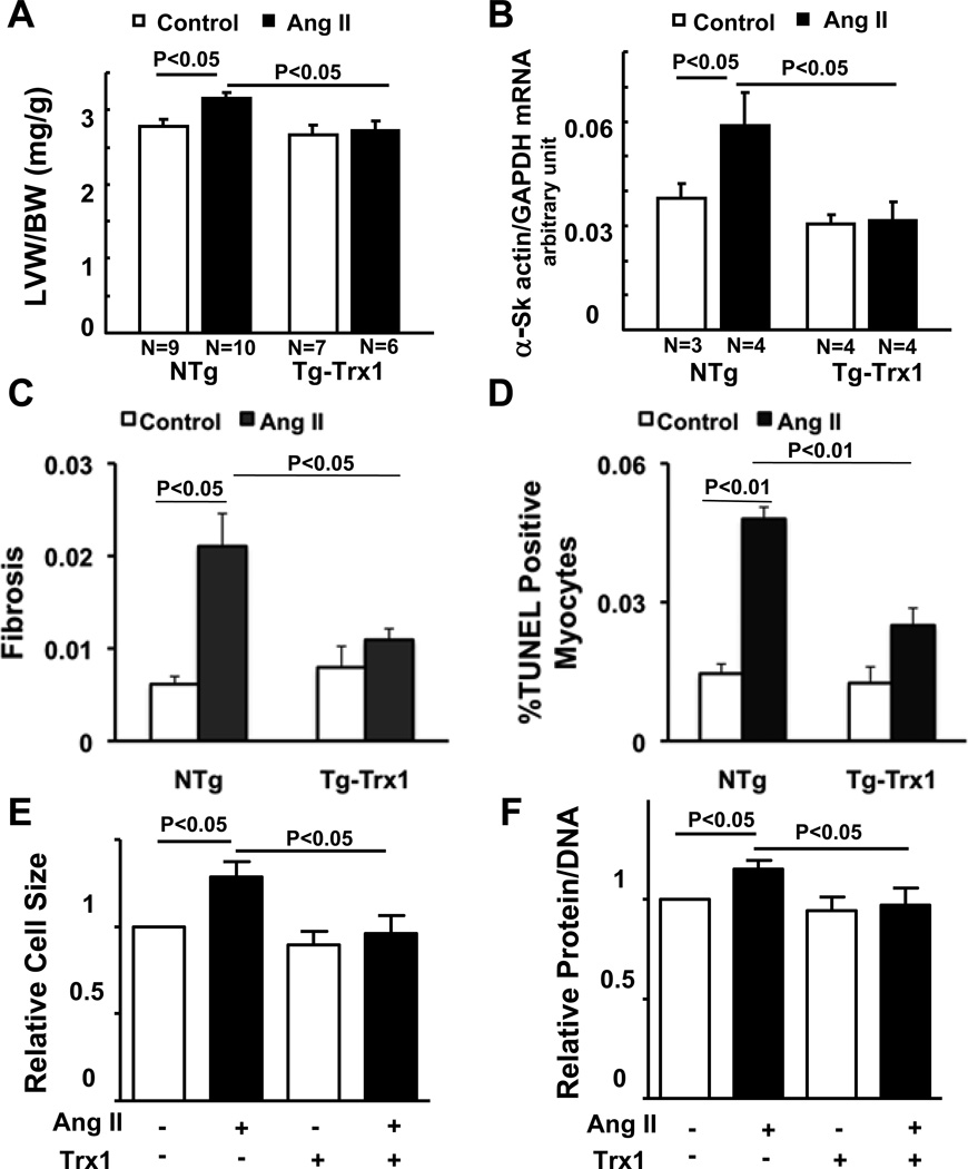
Figure 1. Trx1 attenuates Angiotensin II (Ang II)-induced cardiac hypertrophy, fibrosis and apoptosis.
(A–D) Tg-Trx1 and NTg mice were subjected to continuous infusion of either PBS or a subpressor dose (200 ng/kg/min) of Ang II by osmotic pumps for two weeks. (A) Postmortem measurements of left ventricular weight/body weight (LVW/BW, mg/g) after two weeks infusion. (B) qRT-PCR analysis of heart homogenates from Tg-Trx1 and NTg mice with or without Ang II infusion. (C) Fibrosis was assessed by periodic acid-Schiff (PASR) staining and the fibrotic area was measured. (D) Cardiomyocyte apoptosis in the mouse hearts was observed using TUNEL staining, and the TUNEL positive cells were counted. (E, F) Twenty-four hours after transduction of Ad-LacZ or Ad-Trx1, neonatal rat cardiomyocytes (NRCMs) were treated with or without 100 nM Ang II for 48 hours. Cells were stained with anti-α-actinin antibody and DAPI, and relative cell surface area (cell size) was measured (E), or were harvested for protein and DNA content measurement (F). N=3. Values are mean±SEM. P<0.05.

