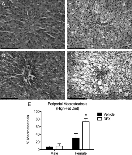
Fig. 3.
Fetal DEX exposure causes elevated periportal steatosis in DEX-exposed females (magnification, ×20). Scale bars, 100 μm. A significant increase in macrosteatotic vesicles was found in livers from female offspring that had been exposed prenatally to DEX (D), but not vehicle (C). This was not observed in the male offspring (A and B). Macrosteatosis in the female liver also resulted in substantial disruption of hepatic architecture (D). Cell counts are reported as mean ± sem of six animals, with significant differences between vehicle and DEX exposed offspring indicated by asterisk (E).

