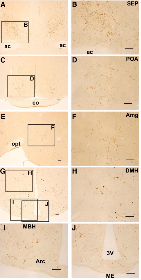
Fig. 4.
Location of GnIH-ir fibers and cell bodies in the hamster brain. A, GnIH-ir fibers in the septal region. ac, Anterior commissure. Bar, 100 μm. B, Higher magnification of the highlighted area in A showing GnIH-ir fibers in the SEP. Bar, 100 μm. C, GnIH-ir fibers in the anterior hypothalamic region. co, Optic chiasm. Bar, 100 μm. D, Higher magnification of the highlighted area in C showing GnIH-ir fibers in the medial POA. Bar, 100 μm. E, GnIH-ir fibers in the amygdaloid region. Medial is to the left. opt, Optic tract. Bar, 100 μm. F, Higher magnification of the highlighted area in E showing GnIH-ir fibers in the medial amygdaloid nucleus (Amg). Bar, 100 μm. G, GnIH-ir fibers in the medial hypothalamic region. Bar, 100 μm. H, Higher magnification of the highlighted area in G showing GnIH-ir fibers in the DMH. Bar, 100 μm. I, Higher magnification of the highlighted area in G showing GnIH-ir fibers in the MBH. Arc, Arcuate nucleus. Bar, 100 μm. J, Higher magnification of the highlighted area in G showing GnIH-ir fibers in the median eminence (ME). 3V, Third ventricle. Bar, 100 μm.

