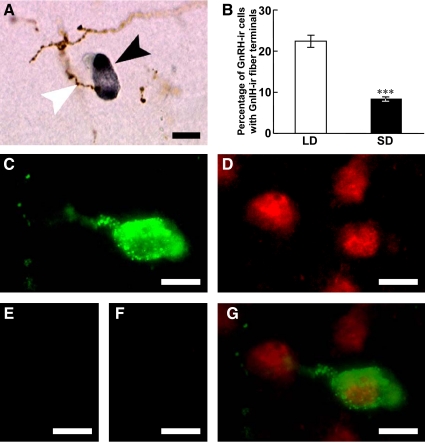
Fig. 7.
Effects of photoperiod on the interaction of GnIH-ir fibers with GnRH-ir neurons and expression of GnIH receptor in a GnRH-ir neuron. A, GnIH-ir fibers (brown) and a GnRH-ir neuron (dark gray) in the medial POA. A white arrowhead indicates a GnIH-ir fiber in close proximity to a GnRH neuron indicated by a black arrowhead. Bar, 10 μm. B, Percentage of GnRH-ir cells with a close apposition of a GnIH-ir fiber terminal in hamsters kept in LD or SD for 13 wk. Each column and the vertical line represent the mean ± sem (n = 4 samples; one sample from one animal). ***, P < 0.001 vs. LD by Student's t test. C, Fluorescence microscopic image of GnRH-ir neuron (green) in the POA. Bar, 10 μm. D, Fluorescence microscopic image of GnIH receptor (red) in the same section. Bar, 10 μm. E, Immunocytochemistry without anti-GnRH antibody showed no fluorescence. Bar, 10 μm. F, Immunocytochemistry without anti-GnIH receptor antibody also showed no fluorescence. Bar, 10 μm. G, Merged image of C and D showed that GnIH receptor (red) was expressed on GnRH-ir (green) neuron. Bar, 10 μm.

